• Dysfunctional T cell signaling is involved in many diseases • Membrane biophysical properties can modify cell signaling • Membrane lipid order is linked to cell signaling in T helper cells
THE immune system works to protect an individual from external and internal threats – such as bacteria, viruses, and cancers – and is composed of two complementary subsystems: innate and adaptive immunity.
The innate immune system is present at birth; provides a rapid, non-specific response to foreign threats; and is composed of physical barriers like skin and mucus, and specialized cell types including monocytes and neutrophils. The adaptive immune system, on the other hand, learns about threats from the innate immune system and develops specific defenses against them, which can be long-lasting; it is composed of specialized cell types called lymphocytes.
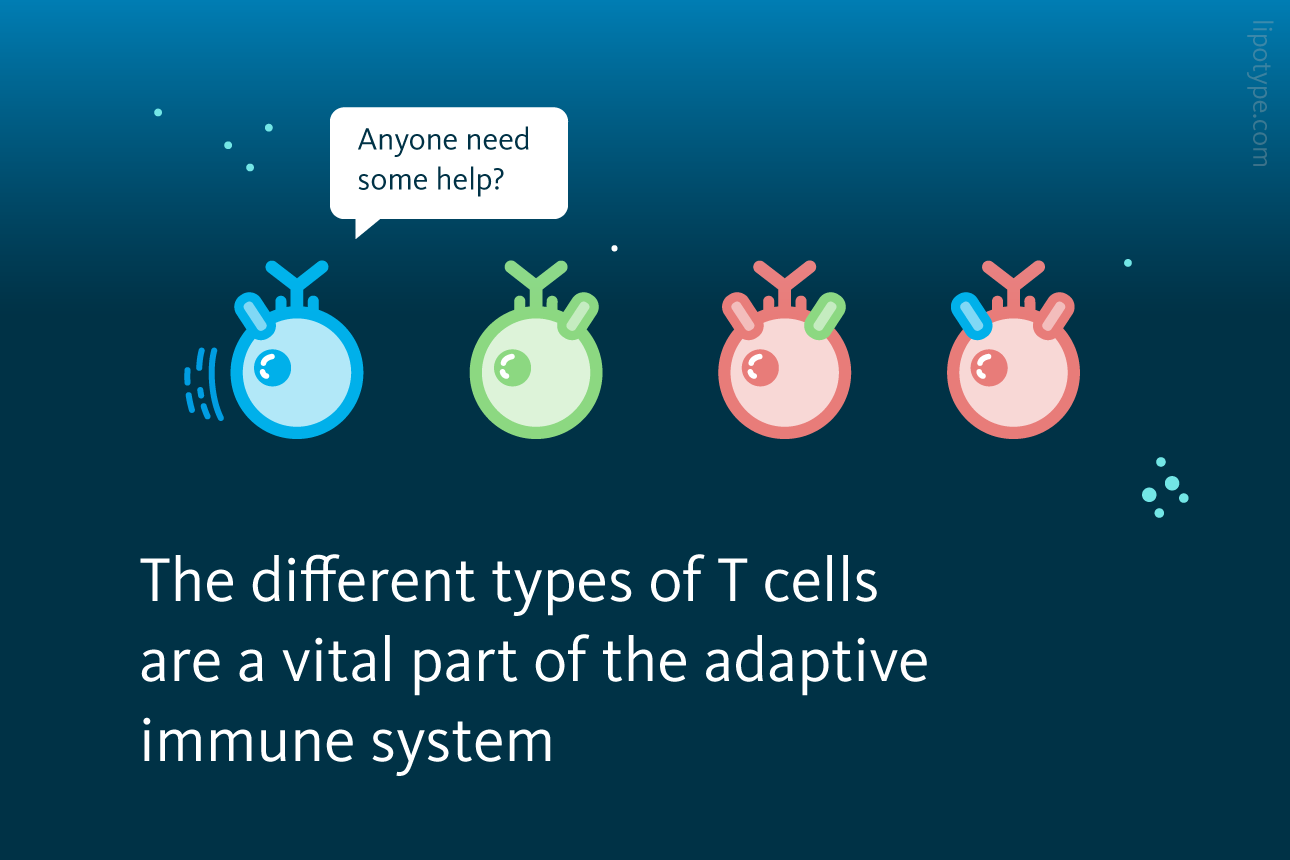
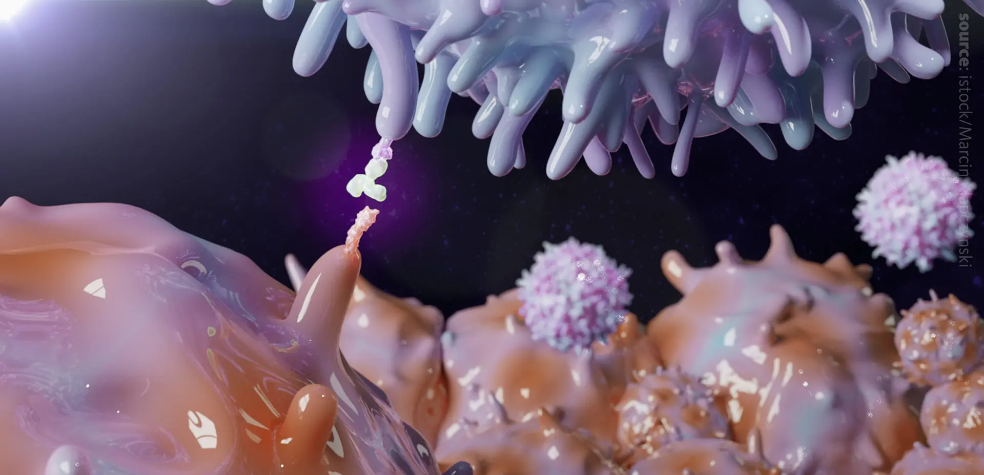
T cells are a subset of lymphocytes that – through unique, surface-bound proteins known as T cell receptors (TCRs) – directly recognize and target threat-associated antigens. T cells also communicate with other cells in the adaptive immune system by producing chemical signals known as cytokines. There are many types of T cells, including: cytotoxic T cells, which directly mediate the killing of infected cells; T helper cells, which support and modulate the activity of other T cells; and T regulatory cells, which dampen the immune response and support distinguishing between self and non-self molecules to reduce the risk of autoimmune diseases.
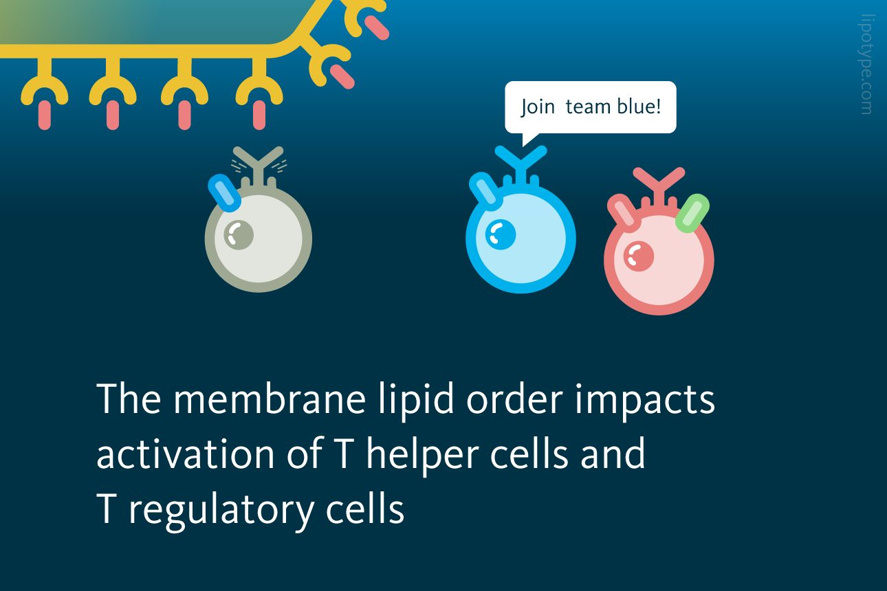
T cell signaling can, like other processes that occur at cellular membranes, be modified by the biophysical properties of membranes. For example, membrane lipid order – a measure of how efficiently lipids can pack together – influences the formation, persistence, and function of signaling platforms. Membrane lipid order depends on membrane composition, environmental temperature, lipid metabolism, and other factors.
Previous work suggests that the liver X receptor (LXR) – a transcription factor that regulates many lipid metabolic processes – can modify the function of T cells, potentially by altering membrane biophysical properties. To further explore these relationships, a London-based team of scientists analyzed the transcriptome, lipidome, and phenotype of T helper cells. The team’s results showed that LXR regulates glycosphingolipid metabolism in T helper cells, which in turn modifies immune signaling and function.
LXR pathways associated with membrane lipid composition: LXR is a nuclear transcription factor that influences the regulation of a wide range of metabolic processes, including lipid and glucose homeostasis. Shown are select enzymes which are regulated by LXR and their relation to membrane composition.
Peripheral blood samples were collected from 50 healthy volunteers. T helper cells were isolated and characterized via transcriptomic, lipidomic, and phenotypic assays. Results were compared with data from other immune cells and from T helper cells following LXR and/or TCR stimulation.
First, control T helper cells and those treated with an LXR agonist were analyzed via RNA-seq; 65 genes were differentially expressed between the two groups. Hierarchal clustering into functional pathways linked the 53 upregulated genes to lipid metabolism and the 12 downregulated genes to inflammation.
Lipid metabolism regulation by LXR in CD4+ T cells: Primary human CD4+ T cells (n = 3) were cultured with or without LXR agonist (GW3965, GW) for 24 h, then, gene expression was assessed by RNA sequencing. These network diagrams illustrate pathways that are significantly enriched for up- or down-regulated genes, where each node represents a significantly altered term and node size proportional to the number of contributing genes. Similar terms with a high degree of redundancy were clustered. Waddington et al., PNAS (2021), doi: 10.1073/pnas.2017394118
Following stimulation of both LXR and TCR, genes involved in lipid metabolism – including of cholesterol – were upregulated; and genes involved in immune processes – including leukocyte activation, chemokine production, and chemotaxis – were downregulated. Additionally, these cells produced more IL-2 and IL-4 and less IL-17A (all three cytokines are involved in T cell activation) and proliferated less.
Next, control and treated T helper cells were analyzed via lipidomics. The total lipid content of cells in the two groups was the same. However, the levels of both triacylglycerols and hexosylceramides increased.
LXR activation changes lipid composition in T helper cells:A Human CD4+ T cells (n = 4) were cultured with LXR agonist (GW) or without it (CTRL) for 36 h. Lipidomics analysis shows change in triacylglycerol (TAG) and hexosylceramide (HexCer) levels relative to total lipids. B Cells were cultured with LXR agonist (GW) or without it (CTRL) for 24 h. CD4+ T cells were selected for filipin staining for cholesterol. Cumulative data is showing the change in geometric mean fluorescence intensity (gMFI). Waddington et al., PNAS (2021), doi: 10.1073/pnas.2017394118
The latter observation was attributed to increased expression of the glycosphingolipid biosynthesis enzyme UDP-glucosylceramide synthase (UGCG), which is the rate-limiting enzyme for the conversion of ceramides to hexosylceramides. UGCG was also shown to be directly regulated by LXR. Simultaneously, plasma membrane levels of cholesterol were reduced, which was attributed to the increased expression of the cholesterol efflux transporters ABCA1 and ABCG1.
Both, hexosylceramides and cholesterol are important components of membrane lipid rafts, subdomains of the plasma membrane which facilitate interactions of membrane proteins. Cholesterol levels correlate positively with T plasma membrane lipid order, glycosphingolipids levels – such as hexosylceramides – correlate negatively with T plasma membrane lipid order. Thus, the transcriptomic and lipidomic data from T helper cells both showed that LXR activation reduces membrane lipid order.
Immune synapse formation is regulated by LXR activation: CD4+ T cells were cultured with LXR agonist (GW) or without it (CTRL) before they were added to chamber slides coated with anti-CD3/28 for immune synapse formation. A Immune synapses were fixed at 15 mins post activation and immunostained for Lck to be quantified by corrected total cell fluorescence. B Band intensities relative to HSP90 (loading control) were calculated for signaling protein phosphorylation after 2, 5, and 10 minutes of T cell receptor engagement by Western blotting. Waddington et al., PNAS (2021), doi: 10.1073/pnas.2017394118
The reduced membrane lipid order modified the immune synapse – which is the location of TCR-mediated cell activation – in T helper cells. Following LXR activation, the receptor tyrosine kinase Lck accumulated at the edges of the immune synapse, which has been linked to T cell activation. Additionally, phosphorylation of two proteins involved in T cell signaling – CD3 and LAT – increased.
Additional experiments showed that, compared to T helper cells, regulatory T cells exhibited lower membrane order and higher glycosphingolipid levels than T helper cells. Cholesterol levels were lowered similarly. Subsequent LXR stimulation in regulatory T cells altered lipid metabolism in ways that were distinct from those observed in T helper cells, suggesting that the LXR pathway may contribute to T cell specialization.
Comparison of LXR activation induced changes in T helper and T regulatory cells: Human CD4+CD25low T helper cells (Thelp) and CD4+CD25+ T regulatory cells (Treg) were cultured with LXR agonist (GW) or without it (CTRL) for 24 h. Lines connect CTRL- and GW-treated samples from the same donor. A Cumulative change in percentage of cells highly expressing cholera toxin B (CTB high %) as a surrogate marker for glycosphingolipids. B Cumulative data of filipin staining for cholesterol is showing the change in geometric mean fluorescence intensity (gMFI). Waddington et al., PNAS (2021), doi: 10.1073/pnas.2017394118
The biophysical properties of membranes – such as lipid order, membrane fluidity, and composition – play an influential role in cell signaling and function. Overall, this study demonstrates that LXR activation alters lipid metabolism and, through mechanisms that involve membrane lipid order and subsequent modification of cell signaling, immune function in T helper cells. This finding indicates that LXR may be an effective therapeutic target for diseases that involve aberrant T cell signaling, such as cancers and autoimmune disorders.
Through characterization of the lipidome of whole cells or cell fractions, Lipotype Lipidomics technology can help elucidate the relationships between membrane biophysical properties, lipid metabolism, and physiological and pathological processes.
University College London (UCL) is a world-leading research university with over 6,000 academic and research staff committed to using their collective expertise to address global problems. UCL is working to extend the human knowledge of the local, national and global.
Share this story
About Lipotype
Lipotype is the leading lipidomics service provider to reach your research goals. Order your lipidomics service, send in your samples and get your data in as little as two weeks.
You are currently viewing a placeholder content from OpenStreetMap. To access the actual content, click the button below. Please note that doing so will share data with third-party providers.


 in the thymus. Both, negative and positive selection are shown.")

 were cultured with or without LXR agonist (GW3965, GW) for 24 h, then, gene expression was assessed by RNA sequencing. These network diagrams illustrate pathways that are significantly enriched for up- or down-regulated genes, where each node represents a significantly altered term and node size proportional to the number of contributing genes. Similar terms with a high degree of redundancy were clustered.")
 were cultured with LXR agonist (GW) or without it (CTRL) for 36 h. Lipidomics analysis shows change in triacylglycerol (TAG) and hexosylceramide (HexCer) levels relative to total lipids. B Cells were cultured with LXR agonist (GW) or without it (CTRL) for 24 h. CD4+ T cells were selected for filipin staining for cholesterol. Cumulative data is showing the change in geometric mean fluorescence intensity (gMFI).")
 or without it (CTRL) before they were added to chamber slides coated with anti-CD3/28 for immune synapse formation. A Immune synapses were fixed at 15 mins post activation and immunostained for Lck to be quantified by corrected total cell fluorescence. B Band intensities relative to HSP90 (loading control) were calculated for signaling protein phosphorylation after 2, 5, and 10 minutes of T cell receptor engagement by Western blotting.")
 and CD4+CD25+ T regulatory cells (Treg) were cultured with LXR agonist (GW) or without it (CTRL) for 24 h. Lines connect CTRL- and GW-treated samples from the same donor. A Cumulative change in percentage of cells highly expressing cholera toxin B (CTB high %) as a surrogate marker for glycosphingolipids. B Cumulative data of filipin staining for cholesterol is showing the change in geometric mean fluorescence intensity (gMFI).")