Regulating fatty acid metabolism using lipases could be beneficial in Parkinson’s disease treatment.
About the author
Olga (Olya) Vvedenskaya Sci. Communications Officer
Dr. Dr. Olya Vvedenskaya studied medicine, and further obtained her PhD in the field of molecular oncology. She loves to deliver scientific messages in a clear and accessible manner.
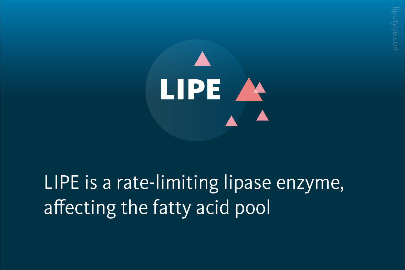
• LIPE is a triacylglycerol lipase • LIPE affects the fatty acid pool • Targeting fatty acid generation through the neutral lipids degradation pathway via LIPE rescues PD phenotype
Olga (Olya) Vvedenskaya Sci. Communications Officer
Dr. Dr. Olya Vvedenskaya studied medicine, and further obtained her PhD in the field of molecular oncology. She loves to deliver scientific messages in a clear and accessible manner.
LIPIDS and fatty acids are important for the development of the brain and the function of neurotransmitters and receptors. Cells control the production and distribution of fatty acids, which are stored as triacylglycerols (TAGs) in small structures called lipid droplets (LDs) to prevent harmful effects of fatty acid accumulation. These droplets are present in most cells but are less common in neurons, indicating that the regulation of fatty acid production and metabolism is even more important in the central nervous system to prevent harmful buildup of free fatty acids.
Parkinson’s disease and other synucleinopathies cause an accumulation of a protein called α-Synuclein, which forms Lewy bodies and Lewy neurites. Lewy bodies are abnormal clusters of α-Synuclein that develop in the brain. As well as containing the disease hallmark α-Synuclein protein, Lewy Bodies also contain lipids.
The protein α-Synuclein is a small molecule found in nerve cells, that is involved in both normal and disease-related processes. It interacts with lipids in cell membranes and can alter the balance of lipids in the cell. Overproduction of α-Synuclein leads to the formation of lipid droplets, an indication of a dysregulated neutral lipids pathway to store disease-associated excess fatty acid. Lipidomics of Parkinson’s Disease versus non-disease controls identifies lipids as altered in Parkinson’s Disease (including brain, CSF, and plasma).
Fanning and Selkoe identified a new therapeutic target, a triacylglycerol lipase, called LIPE, which affects lipid metabolism by breaking down TAGs. They found that LIPE plays a role in regulating the levels of phospholipid-incorporated unsaturated fatty acid content. This is important because changes in the composition of these lipids can affect how α-Synuclein interacts with cell membranes. By targeting LIPE, it may be possible to modify these interactions and potentially treat Parkinson’s disease.
A schematic representations of neutral lipid synthesis and degradation pathways. Red frames indicate lipid classes: G, glycerol; DAG, diacylglycerols; TAG, triacylglycerol; MAG, monoacylglycerol. Green frames indicate lipases: ATGL, adipose triglyceride lipase; LIPE, hormone-sensitive lipase; MGL, monoglyceride lipase. FA, fatty acid; LD, lipid droplet; * points to rate-limiting step, dashed line goes for upstream synthesis pathway. Fanning et al., npj Parkinson’s Disease (2022) 74, 10.1038/s41531-022-00335-6.
Reducing the metabolic activity of LIPE both genetically and pharmacologically has been found to decrease the formation of abnormal accumulation of α-Synuclein in cells, and a decrease in the unfolded protein response. Specifically, the decrease in lipase activity was associated with a decrease in the levels of certain fatty acids, including 18:1n9, 16:1n9, 16:0, and 18:1n7. The decrease in 18:1n9 was particularly noteworthy because it is a highly abundant fatty acid.
Regulation of fatty acid composition with LIPE inhibition. Upper image: LIPE knockdown decreases monounsaturated FAs. Lower image: LIPE pharmacological inhibition decreases monounsaturated FAs in α-Synuclein-expressing cells. Red and blue heatmap is a representation of a given FA species relative amount. Saturated/unsaturated status indicated by white/black squares. Fanning et al., npj Parkinson’s Disease (2022) 74, 10.1038/s41531-022-00335-6.
The findings from both genetic and pharmacological experiments indicate that reducing the LIPE activity can lower levels of unsaturated fatty acids in phospholipids and reduce the accumulation of α-Synuclein at membranes in cells. These results suggest that slowing down the lipid degradation process by targeting LIPE may be a promising approach for developing new therapies for Parkinson’s disease.
In experiments using Caenorhabditis elegans, a type of worm used as a model organism, reducing LIPE activity rescued the loss of dopaminergic neurons caused by α-Synuclein. In primary cell culture experiments using neurons from PD patients with α-Synuclein triplication (essentially, neurons with Parkinson’s disease phenotype), LIPE inhibition reversed abnormal levels of phosphorylated α-Synuclein and the unfolded protein response, as well as increasing the ratio of tetrameric to monomeric α-Synuclein. LIPE inhibition also restored the phospholipid-incorporated fatty acid profile of α-Synuclein triplication neurons to that of healthy neurons.
LIPE inhibitor effect on the primary neurons. LIPE inhibitor restores the fatty acid composition of α-Synuclein triplication neurons to a normal state, similar to control neurons. Red and blue heatmap is a representation of a given FA species relative amount. Saturated/unsaturated status indicated by white/black squares. Fanning et al., npj Parkinson’s Disease (2022) 74, 10.1038/s41531-022-00335-6.
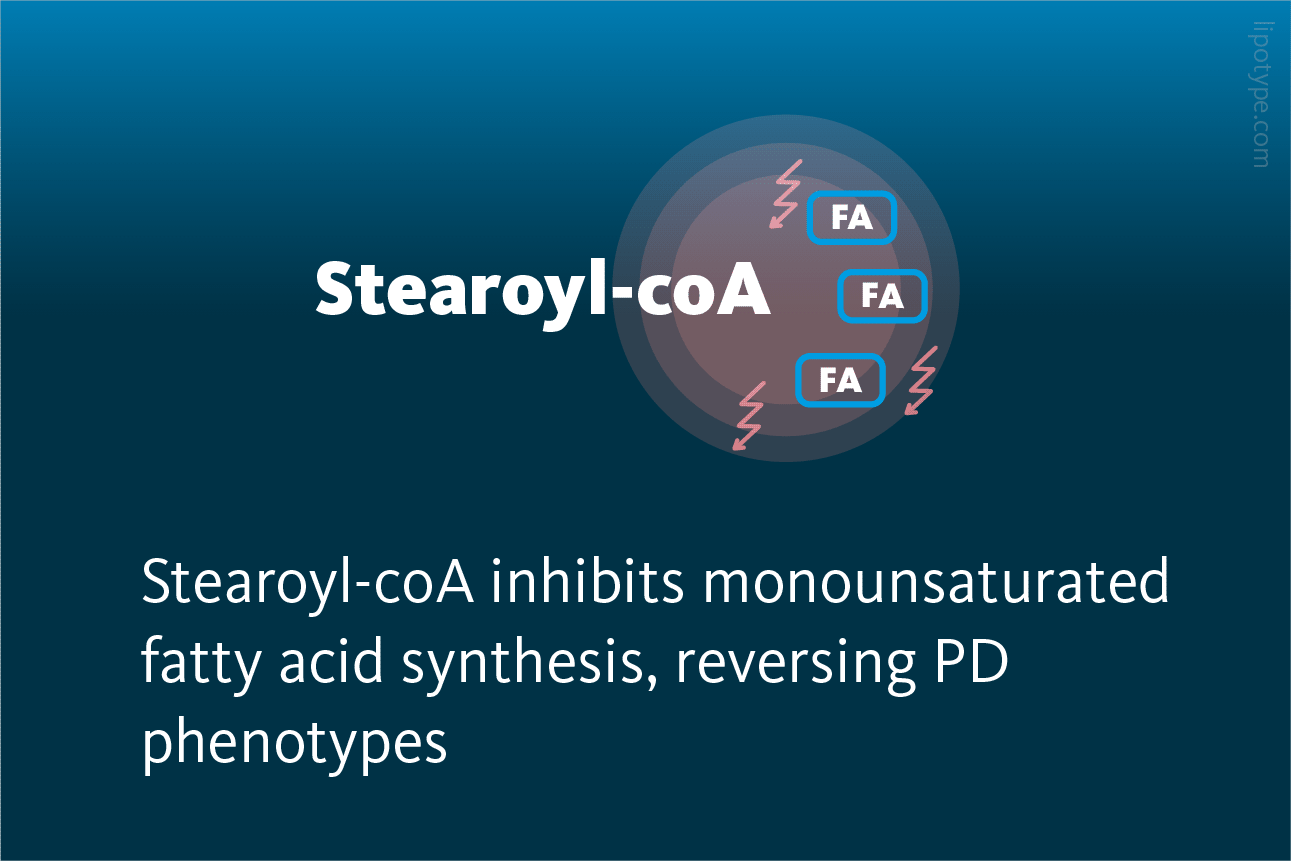
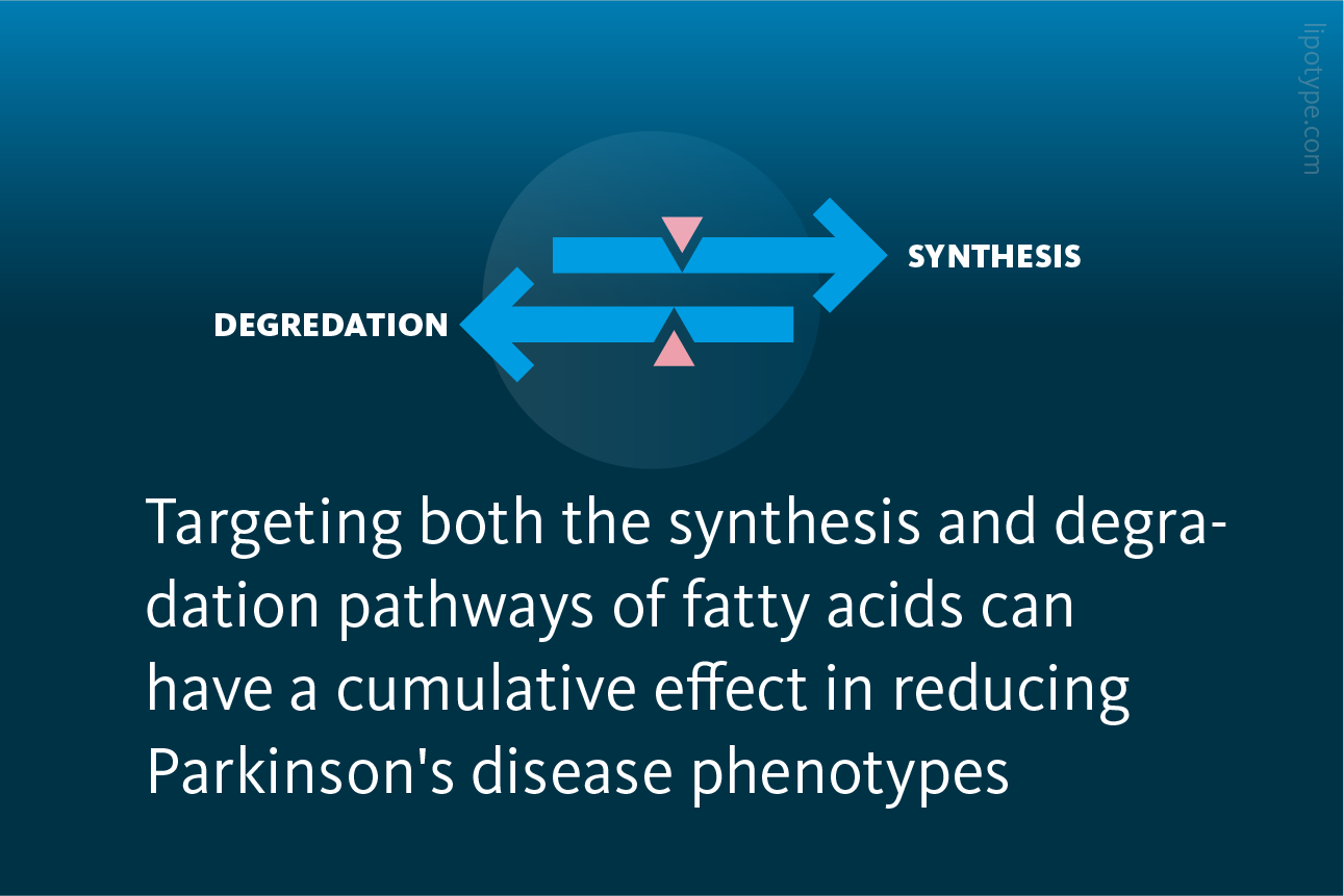
Targeting fatty acid levels through both synthesis and degradation pathways by targeting a desaturase (SCD) and a lipase (LIPE) can lead to additive effects, resulting in reduced α-Synuclein inclusions and phosphorylation. Together, these interventions cumulatively decrease 18:1n9 and 16:1n9 levels, leading to decreased α-Synuclein inclusion formation and phosphorylation in patient-derived α-Synuclein triplication neurons.
LIPE inhibition reduces 16:1-containing fatty acid species in several phospholipid classes. PS, PE, PC, PI classes containing 16:1 species are decreased upon LIPE inhibition. Middle line: mean values. Error bars: standard deviation (n in graph order: 5, 6). **p < 0.01 unpaired t-test. PS, phosphatidylserine; PE, hosphatidylethanolamine; PC, hosphatidylcholine; PI, phosphatidylinositol. Fanning et al., npj Parkinson’s Disease (2022) 74, 10.1038/s41531-022-00335-6.
α-Synuclein protein has been shown to interact with membranes, and alterations in membrane composition can impact α-Synuclein-membrane interactions, leading to aggregation and toxicity. This study identified LIPE as a potential therapeutic target for modifying synucleinopathy phenotypes. LIPE regulates phospholipid-incorporated fatty acid content, particularly unsaturated fatty acid levels, which are important in α-Synuclein:membrane interactions.
Reducing LIPE activity decreased α-Synuclein accumulation in round, membrane-rich cytoplasmic inclusions and decreased PD-associated phosphorylated α-Synuclein and insoluble α-Synuclein levels. LIPE inhibition also increased the abnormally low α-Synuclein tetramer to monomer ratio and increased the ratio of cytosolic to membrane-bound α-Synuclein in patient-derived α-Synuclein triplication neurons. Therefore, targeting fatty acid metabolism through the lipid degradation pathway, particularly through the modulation of phospholipid-incorporated fatty acids, represents a promising strategy for treating synucleinopathies.
Harvard Medical School is a leading institution in medical education, research, and clinical care. The research focus of Harvard Medical School is broad and covers a wide range of fields within the biomedical sciences such as cancer biology, neuroscience, genetics and genomics, infectious diseases, immunology, and stem cell biology.
Newsletter
Get the latest lipidomics articles and publications delivered straight to you.
You are currently viewing a placeholder content from OpenStreetMap. To access the actual content, click the button below. Please note that doing so will share data with third-party providers.


. **p < 0.01 unpaired t-test. PS, phosphatidylserine; PE, hosphatidylethanolamine; PC, hosphatidylcholine; PI, phosphatidylinositol.")