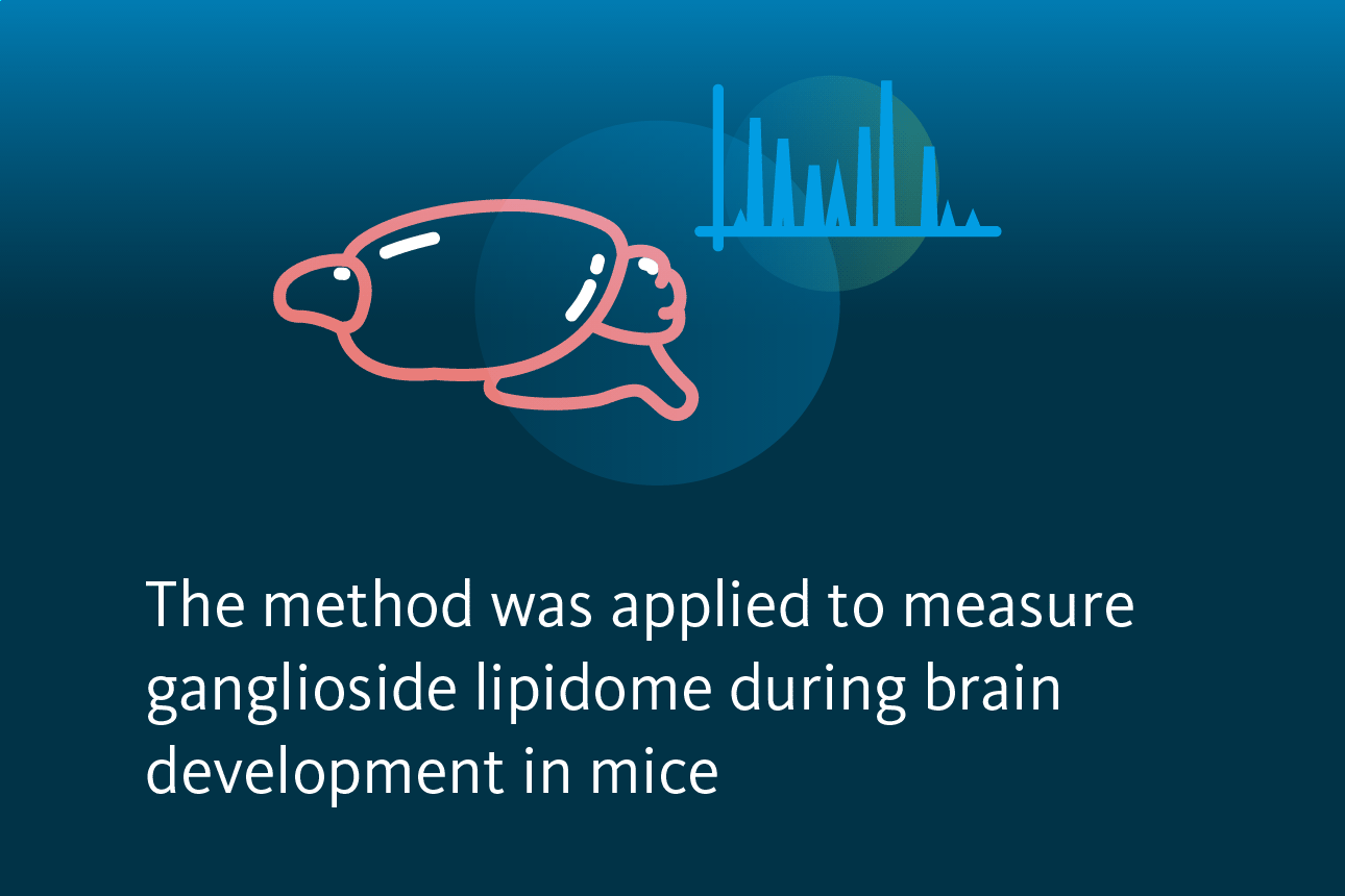
The central nervous system (CNS) and peripheral nervous system are composed of cells capable of electrical excitability, such as neurons, as well as non-excitable cells that offer essential support, known as glia cells. Within the central nervous system, glia cells consist of oligodendrocytes, astrocytes, and microglia. Oligodendrocytes primarily function in producing myelin, a multi-layered membrane that provides insulation for neuronal axons. Axons contain concentrated gangliosides, playing a role in axon-myelin interactions. However, the alterations in ganglioside composition during myelination remain unclear. In an effort to address this gap, a group of researchers, including scientists from Lipotype, teamed up to analyze the ganglioside lipidome in mice throughout development and adulthood. Additionally, the team investigated the ganglioside content in mice lacking genes responsible for synthesizing the majority of ganglioside species.
Gangliosides exhibit significant structural heterogeneity, forming a diverse group of ganglioside species. The synthesis of gangliosides is a regulated, region-dependent, and sequential process. Initially, glycosphingolipid precursors are produced in the endoplasmic reticulum, followed by the glycosylation of ceramide in the Golgi apparatus. This process results in the formation of glucosylceramide (GlcCer), which is subsequently converted into lactosylceramide (LacCer), serving as the metabolic precursor for the synthesis of complex glycosphingolipids.

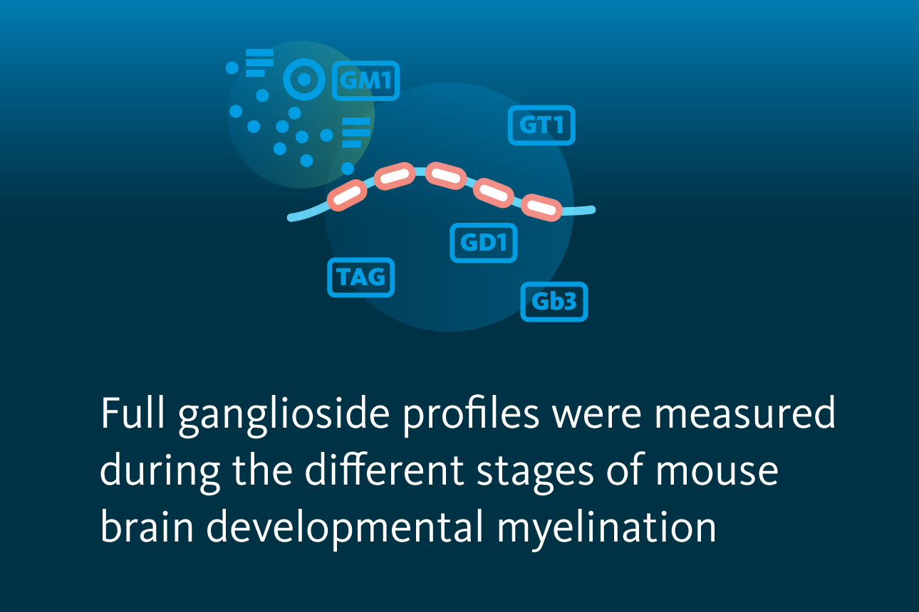
Synthesis of gangliosides.Gangliosides are synthesized from ceramide through the addition of sugar groups and sialic acid as shown on the pathway scheme. Ganlioside synthesis is impaired in mice lacking genes responsible for synthesizing most ganglioside species. These species are highlighted in grey. The main brain gangliosides are highlighted in red.
Arends et al., iScience 2022, 25 (11), 105323, 10.1016/j.isci.2022.105323
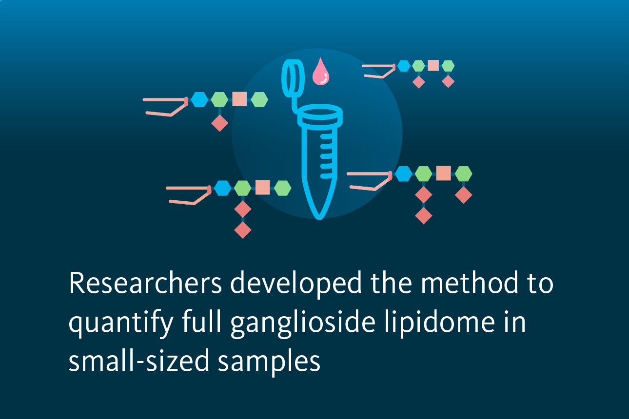
The researchers conducted an analysis of mouse CNS, in particular brain and optic nerve, samples to investigate the role of gangliosides in myelination. In order to facilitate this study, they developed a mass spectrometry analysis technique that enables the quantification of gangliosides in small tissue samples during myelination. Traditional quantitative analysis of ganglioside lipids is typically carried out using liquid chromatography-mass spectrometry (LC-MS), which offers satisfactory sensitivity and reproducibility. Furthermore, LC-MS methods can differentiate between isomeric ganglioside species. However, these approaches demand relatively large sample quantities and involve specialized, often lengthy extraction and enrichment procedures.
To illustrate, the conventional analysis of mouse brain homogenate requires approximately 0.5 g of wet tissue, with extraction performed in 15-20 mL of organic solvents. Both the sample quantity and extraction volume associated with this method make it impractical for high-throughput lipidomic analysis of gangliosides. Moreover, the necessity for substantial sample amounts hinders the examination of small anatomical structures, such as the mouse optic nerve. To address these challenges, scientists from Lipotype, in collaboration with partners, introduced a novel direct infusion (shotgun) lipidomics method that significantly reduces sample requirements to a few hundred milligrams of tissue and extraction volumes below 2 mL.
 as well as polar lipids (mainly phospholipids). From the remaining water phase, the acidic glycosphingolipids (gangliosides) were subsequently enriched based on reverse-phase solid-phase extraction. Gangliosides were eluted and infused into the mass spectrometer with a robotic nano-electrospray ionization device.")
In order to evaluate alterations in lipid composition during CNS developmental myelination, the researchers examined mouse brains at different stages: an early myelination stage, a stage characterized by ongoing but nearly completed myelination, and an adult stage (6 months) when developmental myelination is fully done.
Utilizing the developed method, the researchers determined ganglioside levels in the mouse CNS, starting from the brain samples throughout developmental myelination. The highest quantities were observed in GD1 species, followed by GT1. Lower amounts were found for GM1 and GQ1, while GD3, GT2, GD2, and GM3 species had the lowest levels. Ganglioside species of lower complexity, such as GD2, GD3, and GT2, were notably abundant at both early myelination and nearly complete myelination developmental stages. In contrast, more complex ganglioside species like GD1 and GT1, particularly those with longer carbon chains and more double bonds, were mostly detected in the adult stage. The most abundant gangliosides measured were GD1 36:1; 2 and GT1 36:1; 2 at the early stage of myelination and the adult age. GD1 36:2; 2 ranked among the most abundant gangliosides at early myelination stage, although its quantity significantly decreased during development.

The alteration in ganglioside content within the murine brain during developmental stages. The composition of gangliosides in brain during early myelination, nearly complete myelination, and adult stages. A sample size of 4 for each age group was used. All data are presented as mean ± standard deviation.
Arends et al., iScience 2022, 25 (11), 105323, 10.1016/j.isci.2022.105323
Further analysis of CNS development was performed with assessing the lipid composition of the optic nerve at early myelination, nearly complete myelination, and the adult stage. To demonstrate the applicability of their newly developed method on small tissue samples, the optic nerves of young mice were utilized. Moreover, as optic nerves primarily comprise axons and cells of the oligodendrocyte lineage, this choice of tissue helps reduce the number of gangliosides to those specifically involved in axo-glial processes.
The researchers evaluated ganglioside levels in the optic nerve during development. Consistent with brain samples, GD1 and GT1 exhibited the highest amounts, with the most abundant species GD1 36:1; 2 and GT1 36:1; 2 during the early myelination stage, and all of which decreased during development. Interestingly, GM3, GD2, GT2, and GQ1 were not found in optic nerves, and the levels of GM1 were considerably lower than in brain samples. In the ganglioside classes, shorter carbon chains and a lower number of double bonds were prevalent at early myelination and nearly complete myelination stages, contrasting with the dominance of species featuring longer carbon chains and/or more double bonds at the adult stage.

The alteration in ganglioside content within the murine optic nerve during developmental stages. The composition of gangliosides in optic nerve during early myelination, nearly complete myelination, and adult stages. A sample size of 4 for each age group was used. All data are presented as mean ± standard deviation.
Arends et al., iScience 2022, 25 (11), 105323, 10.1016/j.isci.2022.105323
Finally, the researchers measured ganglioside content of CNS in mice lacking genes responsible for synthesizing most ganglioside species. Their findings confirmed the nearly total absence of gangliosides in the brains of these mice. Interestingly, overall lipid composition of these mice showed no significant difference from that of control mice. To understand how these animals compensate for the absence of gangliosides, the researchers analyzed the length and saturation of the carbon chains. Intriguingly, the average length and saturation of the carbon chains in other lipid classes exhibited minimal changes.
In summary, the researchers successfully developed and applied a quantitative mass spectrometry-based shotgun lipidomics method specifically tailored for analyzing gangliosides. The compatibility of this shotgun method with broader global lipidomic profiling techniques represents a significant technical advancement, unlocking a previously inaccessible area of research.

Lipotype Lipidomics technology is crucial for studying normal myelination, disease-related remyelination, and lipid alterations in neurodegenerative disorders like Alzheimer’s disease, Parkinson’s disease, and multiple sclerosis. It aids in identifying lipid profiles during myelination stages, offering insights into neural repair processes. Identifying lipid biomarkers enables early disease detection and enhances our understanding of neurodegenerative mechanisms, facilitating the development of effective therapies.
Need clarity on the process?
Ask us anything!

Lipotype products are provided for Research Use Only. They are not intended for clinical diagnostic purposes and must not be used to inform medical treatment decisions. The content of this article is for scientific and educational purposes only and should not be considered medical advice.